Mesothelioma
What is mesothelioma?
Mesothelioma is a rare form of cancer that arises from a type of cells known as mesothelial cells that line the body cavities. More than 70 percent of diagnosed cases are pleural (lining of the chest cavity), 20 percent peritoneal (lining of the abdominal cavity), 10 percent pericardial (the sac surrounding the heart) and less than one percent testicular. It usually begins as discrete plaques and nodules that coalesce to produce a sheet-like neoplasm.
Mesotheliomas are classically divided into three primary histologic subtypes with distinct morphology and molecular properties:
- Epithelioid tumors account for 80 to 90 percent of diagnosed cases and have a better prognosis.
- Sarcomatoid tumors give rise to bulky and aggressive masses with the worst prognosis and comprise 10 to 20 percent of diagnosed cases.
- Biphasic tumors are rare and are composed of a mix of epithelioid and sarcomatous tissue.
What causes mesothelioma?
The only known carcinogen implicated in the pathogenesis of pleural mesothelioma is asbestos exposure, which is identified in at least 80 percent of cases. Asbestos is in the environment and patients may not truly be aware that they have been exposed to it. There are some studies indicating that ionizing radiation to the chest and testicular area, and some other minerals such as erionite (similar to amphibole asbestos but only found in countries such as Turkey) may lead to the development of mesothelioma; although these are quite rare.
Because not everyone exposed to asbestos develops mesothelioma, there is growing interest in finding genetic mutations responsible for disease susceptibility. Mutations in the BAP1 tumor suppressor gene have been reported, among others.
Secondary mesothelioma malignancies may also develop after radiation therapy treatment of lymphomas, breast and testicular cancers.
What is asbestos?
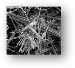
Asbestos is a group of minerals that occur naturally in the environment as bundles of fibers that can be separated to form thin, durable threads.Asbestos fibers have unique qualities:
- They are resistant to heat, fire and chemical degradation; and
- They do not conduct electricity, which made them a desirable substance in many industries.
There are six types of asbestos that may be divided into two groups:
- Serpentine asbestos are long, curly fibers—chrysotile—also known as white asbestos—accounts for 95 percent of all asbestos used worldwide.

- Amphibole asbestos are straight, brittle with needle-like fibers and are more carcinogenic—crocidolite (blue asbestos), actinolite, tremolite, anthophyllite and amosite (brown asbestos).

How does asbestos cause pleural mesothelioma?
The asbestos fibers are inhaled; two-thirds of which are removed through coughing, and the rest enter the distal airways. In the lung, macrophages (a type of white blood cell) attempt to ingest and destroy the remaining fibers. However, the indestructible nature of these fibers resists degradation. Eventually, some fibers pass through the lung and into the pleural space, where the asbestos fibers come in contact with the pleura. In the process, chemical substances are released that cause a slow inflammatory and mutagenic response, as there is an uncontrolled production of macrophages and mutation of the mesothelial cells.
Who is at risk for mesothelioma?
In short: Anyone exposed to asbestos.
Environments and industries with high-exposure to asbestos include:
- Automotive (brakes, insulation and clutches)
- Cement manufacture
- Ceramics
- Construction and demolition (insulation, joint compound, siding, roofing, ceiling and floor tiles)
- Mining
- Paper milling
- Shipyards
- Steam locomotive (insulated pipes and boilers)
Although many patients who develop mesothelioma have had a heavy asbestos exposure over many years, it is not uncommon for patients with a short duration of exposure (i.e., a summer job of one to three months) to develop mesothelioma many years later. Indirect exposure—such as family members who wash the clothing of the workers—can also lead to the development of mesothelioma.
There is no clear association between cigarette smoking and pleural mesothelioma. However, cigarette smokers who have been exposed to asbestos have a higher risk of lung cancer.
What is the incidence of mesothelioma?
Pleural mesothelioma was first reported in 1908, and an association with exposure to asbestos was first proposed in 1960. By 1975, the incidence of pleural mesothelioma began to rise in the U.S. and peaked in 1995, coinciding with diminishing occupational asbestos exposure. Currently, approximately 3,200 cases are diagnosed annually in the U.S. It is more common in men than in women, with a male-to-female ratio of 3:1.
The latency period of mesothelioma—the time elapsed between first exposure to asbestos and diagnosis of the disease—is long and reported at 30-40 years. The approximate age of onset is between 50-70 years old. Approximately two percent of mesothelioma patients are young (< 40 years old) comprising a group with unique clinical characteristics, such as improved overall survival.
What are the signs and symptoms of pleural mesothelioma?
The onset of symptoms is insidious and not specific; typically, once they manifest, the disease is advanced. The most common presenting symptoms of pleural mesothelioma are:
- Dyspnea (shortness of breath) that worsens over time and progresses to shortness of breath at rest. The dyspnea may be a result of pleural fluid accumulation or lung encasement by a tumor.
- Chest wall pain that is usually dull, aching and unrelenting. It is non-pleuritic, meaning it is felt continuously with no variation with respiration.
- Pleural effusion, which is fluid in the pleural space (space between the visceral and parietal pleura).
How is pleural mesothelioma diagnosed?
The diagnosis can be challenging and includes a combination of comprehensive history, physical exam and radiologic findings on a computed tomography (CT) scan of the chest. Tissue biopsy is generally preferred and can be done by several methods, such as a percutaneous needle biopsy. However, this procedure has a low yield for establishing a diagnosis as it rarely provides adequate tissue.
A definitive diagnosis is confirmed based on histologic features and the use of immunohistochemical (IHC) panels. Microscopic evaluation of pleural fluid (cytologic analysis) in search of malignant (cancerous) cells is also an option, as commonly patients present with a pleural effusion. However, it is usually not diagnostic.
What are the treatment options for pleural mesothelioma?
Curative therapy for pleural mesothelioma remains elusive. Patients who are candidates for platinum-based chemotherapy combinations have a modestly-improved survival. There is considerable controversy regarding aggressive surgical approaches and multimodality therapy as there is no clear evidence of the magnitude of benefit. Current studies in immunotherapy (still in its early stages) provide hope to the development of novel approaches to the treatment and prognosis of the disease.
What is the role of VATS in the management of pleural mesothelioma?
Video-assisted thoracic surgery (VATS) is a minimally invasive approach to the chest cavity that allows unparalleled visualization in the chest. It allows for safe removal of a large amount of pleura for definitive diagnosis, and it is virtually 100 percent diagnostic.
Accurate tissue diagnosis of mesothelioma is important to determine:
- If it is mesothelioma or a different tumor that has spread to the lining of the chest
- The type of mesothelioma: epithelial vs. sarcomatoid vs. mixed
- The extent and stage of the disease
- A treatment plan
In addition to obtaining a definitive diagnosis, VATS provides a beneficial therapeutic option: VATS Pleurectomy and talc sclerosis. This is the surgical removal of the parietal pleura with instillation of talc in the pleural space. This procedure has proven very successful at preventing pleural fluid from re-accumulating, thus reducing or eliminating shortness of breath. It also debulks pleural thickening, thus improving pain. It is not a curative measure; it is palliative, but very valuable at improving quality of life.
At times, patients have disease involving the chest wall and visceral pleura causing a trapped lung, meaning the lung cannot re-expand and therefore fluid will re-accumulate. The next best option is insertion of a PleurX catheter through a small incision in your skin into the pleural space. It has many holes to allow the fluid to drain and has a one-way valve that allows the fluid to come out, but does not allow any air to go in. The PleurX catheter can remain in place for weeks or months and is typically drained two to three times per week, allowing patients to breathe comfortably.
See the Mesothelioma Specialists
As the pioneers of video-assisted thoracic surgery (VATS), our experts have decades of experience in minimally invasive surgical treatment and can provide our patients with helpful information on their conditions. Thoracic Group surgeons work in collaboration with a team of specialists, including oncologists, pulmonologists, radiologists, pathologists and nurse navigators to provide the most comprehensive care.
For more information or to schedule an appointment, contact Thoracic Group today.
